Bone marrow film
General Information:
The bone marrow film as well as the blood film are cytological preparations. This is in contrast to the bone marrow biopsy, which is a histological preparation. The normal tissue structure is not present in cytological preparations and many cells are damaged during the preparation.
Assessment:
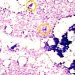
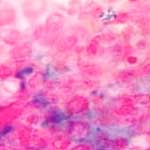
First, bone marrow is examined at 100-fold magnification (10x ocular). The cellularity is age dependent. As a rule of thumb, you can calculate cellularity as 100% minus age. The rest consists of fat vacuoles. In addition, focal cell accumulations, such as lymphoid follicles, can be identified. The dark areas correspond to bone marrow spicules. The megakaryocytes are recognizable at this magnification (see yellow rings in image on the left).
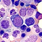
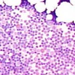
At 400-fold magnification, most cells are identifiable. Normally, a good overall picture can be established. The ratio of myelopoiesis to erythropoiesis is approximately 3-4 to 1. In the bone marrow spicules, the cells are very densely packed and can therefore not be assessed. Only the mast cells are prominent and are seen as dark spots.
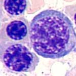
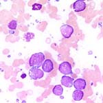
At 1000-fold magnification, the features of the individual cell types can be easily recognized. Damaged cells should not be assessed.
Touch preparation or contact slide:
Bone marrow cannot always be aspirated during an aspiration (dry tap). In such cases, a touch preparation is made. Bone marrow biopsy samples, extracted with a biopsy needle, are lightly pressed against an object slide. The touch preparation is not equivalent to an aspiration preparation. However, it may allow for cytological assessment.
The iron content of bone marrow can be determined in specially-stained bone marrow films (Prussian blue stains).
Bone marrow is also a lymphatic organ. It is the location of primary lymphopoiesis, on the one hand, and the maturation location of the B-cell lymphocytes, on the other hand. Lymphocytes and plasma cells are usually uniformly distributed throughout the bone marrow. However, dense collections of small lymphocytes can also be observed, which usually represent benign lymphoid follicles which are frequently seen in older patients.