May-Grünwald-Giemsa stain (MGG)
Abstract:
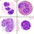
May-Grünwald-Giemsa staining is the stain usually employed for blood and bone marrow films. It consists of a mixture of methylene blue (which dyes acidic cell components blue), azure (which dyes basic cell components red and violet) and eosine (which dyes alkaline cell components orange-red). Since all cell components are stained, this stain is also referred to as panoptic. Since the pH-value is important for stain properties, a pH shift of the solution leads to an incorrect stain reaction. The optimal pH range is from 6.5-6.8.
May-Grünwald-Giemsa is the standard staining method for blood and bone marrow films in Europe. In the United States, the Wright stain is more commonly used. The Wright stain contains eosine and methylene blue dyes and is first heated, in order to make it polychromatic. Since both acidic and basic components of the cells are stained, both staining methods are referred to as panoptic.
Assessment:
In May Grünwald Giemsa stains residual RNA, cytoplasm and nucleoli are blue, DNA and primary granules red and violet as well as hemoglobin and eosinophilic granules are orange-red.
Indication:
All blood and bone marrow films.