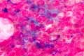
 Hemosiderin stains blue. The iron content of the bone marrow can be determined by the iron stain, which is a good parameter of the body's iron content. Normally, hemosiderin deposits (sideroblasts) are found in a maximum of 4% of the normoblasts. In disorders of hemesynthesis, hemosiderin accumulates in the ring-shaped mitochondria arranged around the nucleus (ringed sideroblasts).
Pappenheimer bodies in the May-Gruenwald-Giemsa stain correspond to hemosiderin occlusions. They typically occur after splenectomy. In the iron stain of the blood film, the Pappenheimer bodies stain blue. Then they are referred to as siderocytes.