 A normochromic, normocytic anemia with anisocytosis, poikilocytosis and tear-shaped erythrocytes as well as a leukoerythroblastic blood picture with normoblasts and early myeloid cells are present. Megakaryocyte remnants are often seen.
Abstract:
Myelofibrosis is a myeloproliferative syndrome and is typified by bone marrow fibrosis. Increasing bone marrow failure results that is compensated for by extramedullary hematopoiesis. The clonal defect is localized in the megakaryocytes. These are increased and produce cytokines and growth factors (among others, platelet derived growth factor = PDGF, platelet factor 4), which results in the proliferation of fibroblasts, and marrow fibrosis.
Clinical picture:
Patients are usually over 50 years of age. The disease begins with fatigue and weight loss. Splenomegaly is obligatory and can lead to abdominal complaints due to enormous size of the spleen. In addition, most patients have hepatomegaly. Under certain circumstances, sclerosis of the bones can be observed by X-ray.
Hematology:
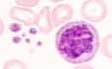
A normochromic, normocytic anemia with anisocytosis, poikilocytosis and tear-shaped erythrocytes as well as a leukoerythroblastic blood picture with normoblasts and early myeloid cells are present. Megakaryocyte remnants are often seen.
Bone marrow:
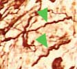 A bone marrow aspiration yields no material ("dry tap"), therefore, a bone marrow biopsy is necessary. The biopsy demonstrates increased megakaryocytes as well as fibrosis. The increase of fibroblasts can be proven by using a silver stain for reticulin fibers (green arrows).