Artifacts
Definition:
Artifacts are blood film abnormalities that occur due to incorrect or faulty processing. These abnormalities make interpretation of the blood film difficult. It is therefore important to recognize these artifacts.
We distinguish the following artifacts:
Blood film is too thin:
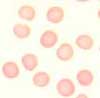
Thin blood films occur when too much pressure or too little blood was used during the preparation of the film. If the film is too thin, the erythrocytes show no central pallor and are often polygonal-shaped. Consequently, the morphology of the erythrocytes cannot be assessed.
Blood film is too thick:
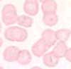
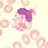
Thick blood films occur when too little pressure or too much blood is applied during the preparation of the film. If the film is too thick, the erythrocytes stack on top of each other and pseudo-rouleaux formation often occurs. This phenomenon is much more frequent in this situation than with multiple myeloma. The morphology of the erythrocytes cannot be assessed.
Staining is too acidic:
The buffer for the Wright staining should have a pH-value between 6.5 and 6.8. If it is too acidic, the erythrocytes may become strongly oxyphilic (acidophilic) and the leukocytes will be poorly stained. Consequently, these leukocytes cannot be assessed.
Staining is too alkaline:
The buffer for the Wright staining should show a pH-value between 6.5 and 6.8. If it is too alkaline, the erythrocytes may become strongly basophilic and the leukocytes demonstrate a toxic appearance with increased granulation and blue cytoplasm.
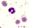
Damaged leukocytes, especially in hyperlipidemia:
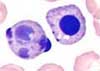
If too much pressure is used when the film is being prepared, the leukocytes could be crushed and can, therefore, no longer be assessed. In the presence of a hyperlipidemia the leukocytes also burst even if the film is correctly prepared. For this reason, carrying out a microscopic leukocyte differential count is not possible. In chronic lymphocytic leukemia (CLL), the leukemic cells are also easily damaged. They are referred to as smudge cells and are a typical feature of CLL.
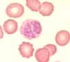
Pseudo-inclusions:

While drying the films, small dots, which look like inclusions in the erythrocytes, can develop. They can easily be confused with Pappenheimer bodies (siderocytes). By adjusting the microscope stage, these pseudo-inclusion become refractile.
Slow processing of blood film:
EDTA blood must be processed for microscopic assessment within 2 hours after the blood has been drawn. If more time elapses, various abnormalities occur in leukocytes. Primarily, vacuoles and coarse granulations occur that cause the blood film to appear toxic. A very typical and unmistakable phenomenon is leukocytolysis. It consists in characteristic abnormalities of the cell nucleus in the form of karyorrhexis and round nuclei. Solid, densely rounded nuclei can sometimes be confused with normoblasts.
Blood stored above room temperature:

If the blood is stored above room temperature after venipuncture, clove-like lobes of the nucleus of lymphocytes may develop.