Plasma cell myeloma
Abstract:
According to the WHO 2016-classification, plasma cell myeloma belongs to the mature B-cell lymphatic neoplasms. A malignant proliferation of plasma cells is present in the bone marrow. The malignant plasma cells usually secrete a monoclonal immunoglobulin of the type IgG (most frequent) or IgA or light chain (M-protein, M=monoclonal, paraprotein). IgD, IgE, or IgM-secreting myeloma are very rare. In 10% of the cases, free light chains are excreted in the urine (Bence-Jones protein). Typically osteolytic lesions and/or diffuse osteoporosis occur. Multiple myeloma constitutes 10-15% of hematological malignancies and usually occurs in older people (mean age = 70 years, rarely under age 40.)
Monoclonal gammopathy of undetermined significance (MGUS) can be a prestage of plasma cell myeloma.
Clinical picture:
Two thirds of the patients complain of joint pain, typically in the back or chest. At the time of diagnosis, osteolysis can be found in 70% of cases by radiography. In advanced stages, pathological fractures can occur. The plasma cell proliferation in the bone marrow causes symptoms of anemia and thrombocytopenia. A tendency toward infection is determined primarily by a decrease in the normal immunoglobulins. Renal failure is mostly determined by the deposition of light chains into the renal tubules. Hypercalcemia can also exacerbate loss of kidney function.
Hematology:
Increased erythrocyte sedimentation rate (ESR) is typical. Rouleau formation of erythrocytes can occur in the blood film. Normochromic-normocytic anemia is usually present. Thrombocytopenia and neutropenia usually reflect the degree of bone marrow infiltration. In rare cases, plasma cells emigrate into the peripheral blood. This is known as plasma cell leukemia.
Bone marrow:

Bone marrow examination, along with serum protein electrophoresis and skeletal surveys, are the main tests for the diagnosis of multiple myeloma.
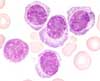
The degree of infiltration can be between a few percent up to over 90% . The plasma cells can have an unobtrusive, mature aspect or look immature with great nucleus-cytoplasm-ratio, fine nuclear chromatin and nucleoli. In immunophenotyping, plasma cells are positive for CD38 and CD138 with expression of cytoplasmic, but not superficial immunoglobulins. In contrast to the wild type-plasma cells, neoplastic plasma cell are mostly negative for CD19, often positive for CD56 and occasionally positive for CD117, CD20, CD10 and CD28 as well as occasionally negative for CD27.
An infiltration of plasma cells of over 30% in the bone marrow with osteolytic lesions or M-protein > 30 g/L is diagnostic. If only a 10-30% marrow plasmacytosis exists, both additional criteria must be fulfilled.
A variety with lymphoplasmacytic aspect of plasma cells typically expresses Cyclin D1 and has the trans-location (11;14).
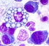
Sometimes round inclusions can be observed in the plasma cells, the so called Russel-bodies. These contain immunoglobulins. They are also observed in reactive processes with intensive production of immunoglobulins. Cells packed with Russel-bodies are called Mott-cells. Inclusions above the nucleus are called Dutcher-bodies. They also contain clonal immunoglobulins.

Flaming plasma cells are a rare phenomenon. They mostly occur in multiple myeloma with an IgA paraproteinemia.
Diagnosis:
According to the WHO classification, a symptomatic plasma cell myeloma can be diagnosed if a paraprotein can be demonstrated in the blood as well as clonal plasma cells in the bone marrow and an impairment of an organ by the myeloma (CRAB-criteria = hypercalcemia, renal failure, anemia, bone lesions). Paraproteinemia and plasma cell infiltration in the bone marrow are not bound to inferior limit. An asymptomatic plasma cell myeloma is present if a paraproteinemia of >30 g/L or a plasma cell infiltration of >10% can be found but no organ symptoms are present (CRAB-criteria negative). The diagnosis of a plasma cell leukemia can be made if more than 20% or > 2.0x10⁹/L clonal plasma cells are found in the peripheral blood.