Peroxidase
Abstract:
Peroxidases occur in numerous body tissues. However, myeloperoxidase only occurs in the primary granules of myeloid cells. It can be detected from the promyelocyte up to the granulocyte stage. Monocytes stain as well, although less positive. Lymphocytic cells are strictly peroxidase-negative.
Assessment:
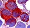 Peroxidase-positive cells demosntrate a reddish-brown granulation. All stages of the myelocytic cell line show a positive reaction with the exception of the myeloblasts. The more mature the cells become, the weaker the reaction becomes. Auer rods yield a strong reaction.
Indication for peroxidase staining:
1. Recognition of acute leukemias of myeloid origin. Caution: A negative reaction makes a lymphocytic origin very probable, however, this does not completely exclude a myeloid origin.
2. A positive result allows for the subdivision of the acute myelocytic leukemias in accordance with FAB classification.
Peroxidase staining is the most important cytochemical investigation for morphological classification of the acute leukemias. Compared to cytochemistry, new investigational techniques such as immunphenotyping, cytogenetics and molecular diagnostic are gaining importance, since they allow for a a much more differentiated and more precise subdivision of the leukemias.
Table of Contents