Bone marrow biopsy
General information:
The bone marrow biopsy is a histological preparation. Unlike the bone marrow film, which is a cytological preparation. In histological preparations, the tissue structure (vessels, fibers, trabecula) is retained. The cytological features of the cells are less readily recognized instead. For this reason, the cells can not always be clearly assigned to a certain type.
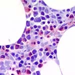
Bone marrow cuts can be stained with hematoxilin-eosin and giemsa. An additional stain is chloracetate esterase with which the myeloid cells stain red (see picture below).
Indication:
A bone marrow biopsy should always be carried out following a bone marrow aspiration. In a bone marrow biopsy, the cellularity can be more easily established than in the bone marrow film. Infiltrative disorders such as metastases, granulomatoses or lymphoproliferative disorders can also be more readily recognized. In parafin-fixied material, the cell types can be more precisely identified immuncytochemically.
Fibrosis:
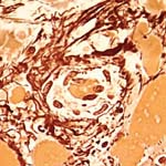
Fibrosis or more precisely, reticulin fiber content, is established in bone marrow cuts stained by silver nitrate. A pronounced increase in fiber content occurs in myeloproliferative syndromes (MPS), and particularly in myelofibrosis.