Hereditary spherocytosis
Abstract:
Spherocytosis is a common autosomal-dominant inherited disease, in which the erythrocytes are spherical due to the heterogeneous defects of various membrane proteins. The erythrocytes are also osmotically more fragile. Spherocytosis is one of the corpuscular hemolytic anemias. Different proteins of the erythrocyte membrane skeleton (e.g. ankyrin, band 3, beta-spectrin) are affected due to genetic defects
The clinical picture ranges from very mild to extremely severe forms.
Clinical picture:
The premature destruction of erythrocytes caused by fragility, known as hemolysis, occurs in the spleen which becomes enlarged. By increasing erythropoiesis, the hematocrit can more or less be maintained normal (compensated hemolysis). Certain infections, especially parvovirus B19, can cause suppression of erythropoiesis. This usually goes unnoticed in patients with normal erythrocytes. However, in patients with hemolytic anemias such as spherocytosis, this can lead to a dramatic decrease of the hematocrit. This phenomenon is called "aplastic crisis" and normally lasts 7-10 days. Various, otherwise trivial infections of the upper air ways or of the gastrointestinal tract can aggravate the hemolytic activity. This is called "hemolytic crisis". Patients often have bilirubin gallstones and cholecystitis due to increased amounts of bilirubin.
Diagnosis and therapy:
The diagnosis is successfully established based on the clinical picture (anemia, icterus, splenomegaly), family history with affected relatives, the hemolytic lab constellation (increased reticulocytes, bilirubin and LDH) and the typical blood film with spherocytes. To confirm the diagnosis, osmotic fragility of red blood cells is measured. It must be increased. Other tests are acid Glycerol-Lysis-test, flow cytometric Eosin-5-Maleinat-binding test and ektacytometry.
Therapy for spherocytosis consists of splenectomy. The microspherocytes' life span clearly increases as a result, however, it does not normalize completely.
Hematology:
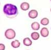
The erythrocytes are round, without central pallor and up to 20-25% are microspherocytes. Hemoglobin concentration is normal to slightly decreased. Reticulocytes are usually increased to over 8%. The MCV is normal to increased, the MCH is normal and the MCHC is increased. The latter occurs only in spherocytosis.
Bone marrow:
An examination of the bone marrow is not necessary for the diagnosis of hereditary spherocytosis. Increased erythropoiesis is present in the bone marrow. Bi-nucleated pronormoblasts and normoblasts can occur.